Welcome to Park Vision’s specialist dry eye clinic
The very latest in ocular surface technology and dry eye research.
Dry Eye Disease (DED), is an increasingly common condition affecting people of all ages. It occurs when there is an insufficient volume and/or quality of tears to keep the surface of the eye sufficiently lubricated.
The common symptoms of DED are: watering, itchiness, burning, redness and irritation. It is a chronic and progressive disease meaning proper diagnosis and management is necessary to address it.
Ocular lubricants (dry eye drops) can be bought easily or prescribed by your GP but they will only temporarily help to manage the symptoms…..and…are you using the right dry eye drops? With so many on the market how do you choose?
Understanding and investigating your dry eye and tear film will allow proper treatment and alleviation of symptoms.
What is tear film?
The tear film is complex containing multiple layers secreted by different glands and tissues. Each layer contains specific molecules and proteins that not only maintain the health of the cells on the ocular surface by providing nourishment and removal of waste products but also protect these cells from environment. The tear film is made up of three layers:
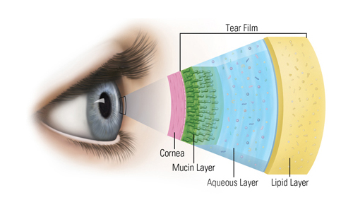
- The mucin layer: this lays against the eye surface and secreted by conjunctival mucous cells. This layer maintains the hydration of the eye, provides lubrication against the conjunctiva and cornea during blink and contributes as a barrier to protect the eye from germs binding to the front surface.
- The aqueous layer: secreted by the lachrymal glands and represents the bulk of the tear film. It is composed of 98% water. The main function of the aqueous layer is to nourish and protect the eye. The aqueous layer carries nutrients and oxygen to the cornea and carries waste products away. It also hydrates the cornea and thereby prevents it from drying out and becoming opaque.
- The lipid layer: secreted by the Meibomian glands. The lipid layer is the outermost layer of the tear film and prevents evaporation of the aqueous layer from the eye. This layer forms a thin, smooth film whose thickness, and structure, influences the rate of evaporation.
Dry eye, put simply, is caused by an impairment of the tear film. This is in the form of insufficient production of tears or the tears evaporating too quickly (evaporative dry eye) and usually caused by a poor or insufficient lipid layer. Evaporative dry eye accounts for 80% of patient sufferers.
At Park Vision we have the latest dry eye technology to investigate, measure and manage your dry eye problems. Offering the latest treatment procedures and leading the field in DED management….
Your Park Vision Dry Eye Assessment
-

Patient history
A complete patient questionnaire and medical history is required to see if medical or environmental factors are contributors.
-
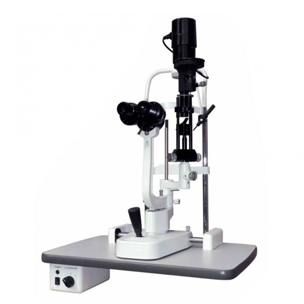
External Examination
An external examination of the anterior eye including lid structure and blink dynamics, using the latest digital slit lamp.
-
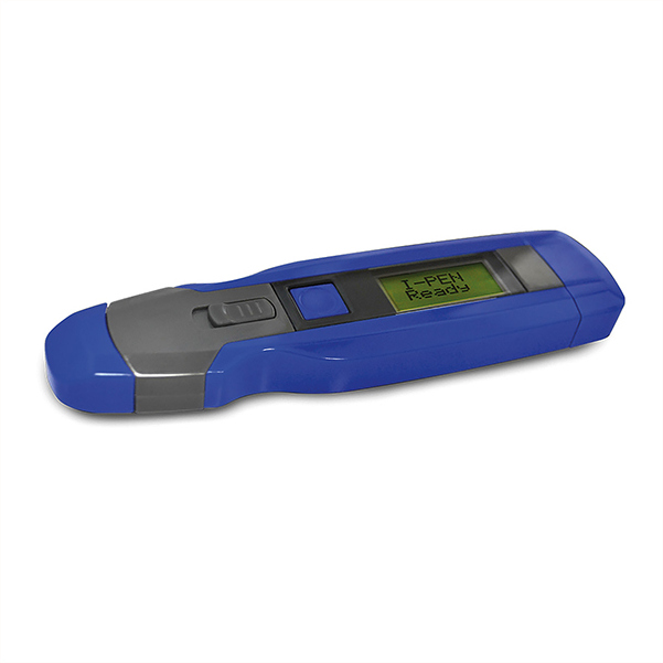
Tear Film Olsolarity
A small tear sample is collected & measured. This helps diagnose ocular surface problems & enables accurate eye-drop prescriptions.
-
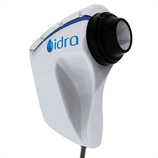
IDRA Tear & Lid Examination
The IDRA is a new and exciting optical instrument that fully measures and assesses the eye-lids and tear film.
The IDRA measures:
- Interferometry: this is a measure of the lipid layer of the tear film and crucial for tear stability. This is recorded and measured against an international grading scale.
- Tear meniscus: the amount of water in the tear oil in millimetres. The IDRA takes a non-invasive photo to observe the quantity, stability and position of the tears against the eye-lid.
- NIBUT: this allows the evaluation of the stability of the mucin layer (the layer closest to the ocular surface), of the tear film and how regularly it breaks down and needs to be refreshed. The automatic grid and non-invasive video record the blink pattern.
- Meibography: using near-infrared light. A completely innovative and revealing picture is taken of the upper and lower eye-lids. This shows Meibomian Gland function – this glands provide meibum, the oily layer required to keep the tear film healthy, stable and the eyes comfortable. Meibomian Gland fall out is a major cause of DED.
- Conjunctival and Corneal staining: the yellow filter on the IDRA and fluorescein on the eye allows the investigation of any staining patterns. This helps classification of the DED condition.